|
|
| .
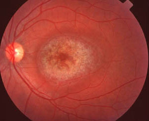
The macula shows an area of hyperpigmentation in the fovea which is surrounded by a zone of depigmentation. The depigmented zone is in turn surrounded by an annulus of hyperpigmentation. The appearance is that of a bull's eye maculaopathy (a better descriptive term is a dartboard appearance but the examiner(s) will probably prefer to hear the conventional description). Further examination:
keratopathy and cataract. common conditions are rheumatoid arthritis (rheumatoid hands, nodules at the elbow) and systemic lupus erythematosus (butterfly rash on the face) (hydroxy)chloroquine toxicity. . |
Questions:
1. What conditions can give rise to bull's eye maculoapthy?