Peripheral corneal thinning / ulceration
|
|||
|
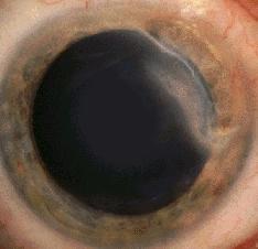
Peripheral corneal thinning can result from inflammation, infection or degeneration. In the examination, the most common cases are Terrien's degeneration, peripheral corneal ulceration secondary to systemic disorders (especially rheumatoid arthritis) and Mooren's ulcer. Although marginal keratitis is the most cause of peripheral thinning and ulceration, it seldom appears in the examination. Terrien's marginal degeneration There are bilateral (but asymmetrical) peripheral thinning
of the superior cornea. The epithelium is intact and contains superificial
vascularization. The thin area has a sloping peripheral border and a sharp
central edge which contains lipid deposits. There may be pseudopterygium
in advanced cases.
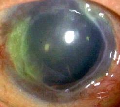
Peripheral corneal ulceration due to systemic diseases There is thinning with ulceration involving a sector of the peripheral cornea. Inflammation may or may not be present (there may be associated scleritis). Look for:
There is (but may be bilateral in younger patient) peripheral
ulcerative keratitis located in the interpalpebral region. The ulceration
is contiguous with the limbus without intervening clear zone. The epithelium
is vascularized and there is an overhanging advancing edge. The whole corneal
circumference may be involved. The sclera is not involved.
|

Questions:
1. Does Terrien's marginal degeneration ever affect the
vision?